
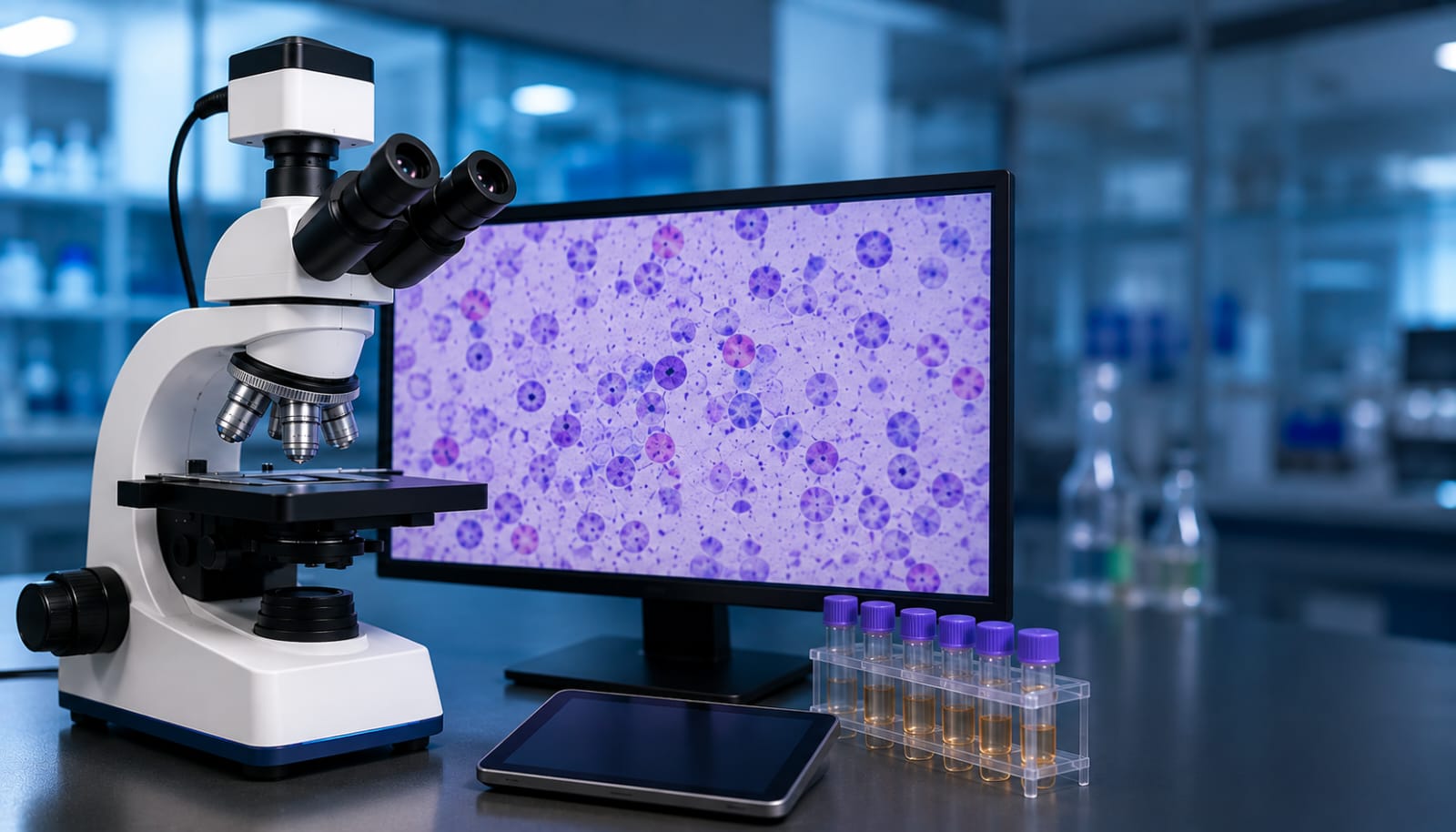
Researchers at the National Institute of Technology (NIT) Calicut have developed an AI-enabled smart microscopy system designed to make medical image analysis faster, more efficient and less demanding for doctors and laboratory professionals.
The technology combines conventional microscopy with artificial intelligence to analyse microscopic images in real time. The system can identify cells that require closer examination, detect abnormalities and highlight other significant features, helping doctors focus their attention on potentially important findings rather than spending long hours manually scanning slides.
One of the key advantages of the system is that it can work with existing microscopes. The AI analysis is performed directly on the device, without depending on an internet connection or cloud-based computing. This makes the technology potentially useful not only in large diagnostic laboratories but also in primary health centres and hospitals where advanced digital infrastructure may be limited.
The system was initially developed for urine sample analysis, with a focus on identifying indications associated with bladder cancer. The researchers believe the technology can eventually be adapted for a wider range of cytology and pathology applications, including blood and liver-related examinations.
The project represents a collaboration between engineering, artificial intelligence and medical expertise. Faculty members and a PhD researcher from NIT Calicut’s Department of Computer Science worked with pathology specialists from Government Medical College, Kozhikode, and KMCT Medical College to develop the AI-assisted microscopy system.
The technology is now being positioned for commercialisation through HealForge AI Private Limited, an NIT Calicut-incubated company. The planned product is intended to bring AI-assisted microscopy into clinical and laboratory environments, potentially supporting healthcare professionals with faster and more efficient image-based analysis.
Funding for the research was provided through the Scheme for Promotion of Academic and Research Collaboration (SPARC), a Government of India initiative under the Ministry of Education.
The research team includes Aarti Menon, PhD researcher in Computer Science at NIT Calicut; Dr Jayaraj P.B., Associate Professor, Computer Science, NIT Calicut; Dr Pournami P.N., Associate Professor, Computer Science, NIT Calicut; Dr Simi S., Assistant Professor of Pathology at Government Medical College, Kozhikode; and Dr Swathi Shankar, Assistant Professor of Pathology at KMCT Medical College, Kozhikode.
With further clinical validation and development, the researchers’ AI-powered microscopy platform could offer a practical route toward bringing advanced image analysis capabilities to a wider range of healthcare facilities.